

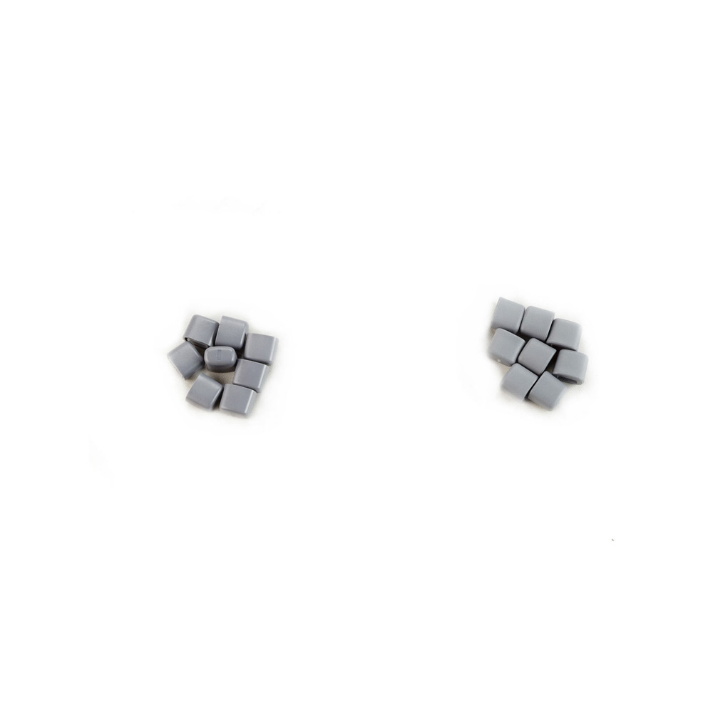
TGC Slide Potentiometer Knob Kit
| 5207000-9 | |
| Ultrasound | |
| GE HealthCare | |
Add to Wish List
Login To Shop
Please select date
Second Opinion Approval Needed
Enter your approval number and submit to add item(s) to cart.
Approval Number
Please enter approval number
OR
Don't know your approval number? Call 800-437-1171
Enter opt 1 for the first three prompts, and have your System ID available.
If you add item(s) to cart and submit your order without the
approval number, GE will contact you before your order
can be confirmed for shipment.
Second Opinion Approval Needed
Select your approver's name and submit to add item(s) to your cart
Approver Name
Please Select Approver Name
OR
Don't know your approval number? Call 800-437-1171
Enter opt 1 for the first three prompts, and have your System ID available.
If you add item(s) to cart and submit your order without
selecting an approver, GE will contact you before your order
can be confirmed for shipment.
Features
- Ingress protection enabled
- Smooth corners and curves
- Sliders gives the entire value range
- Dimensionally stable
Product Overview
The Time Gain Compensation (TGC) slide potentiometer knob kit is used in the ultrasound machines, Magnetic Resonance (MR) machines and also in a few other Digital Imaging (DI) machines. The kit consists of a two bags of slide pot knobs and an instruction sheet. The bag ‘A’ contains 8 slide pot knobs that are compatible with the ALPHA brand slide pots used in the earlier lower OP panel. The bag ‘B’ contains 8 slide pot knobs that are compatible with BOURNS brand slide pots used in the later OP panels. The instruction sheet will have the guidelines and a pictorial representation of the knobs and procedures for using it. The knobs will be fixed to the operator panel and it will have sliding motion to change various parameters and other setups. They are used to compensate for depth related attenuation in an image. The assembled kit will be bagged then each individually boxed and labeled for shipment. It is widely being used in machines such as Optima MR450W 1.5T, LOGIQ E9/S8/200 PRO/9, Voluson E/E8/E8 Expert, Vivid E9/E9 Expert, Precision 500D and other medical diagnostic devices.
Compatible Products

LOGIQ E9
The LOGIQ* E9 is a versatile general imaging system that helps meet a wide variety of general imaging needs. As healthcare evolves and ultrasound exams become more in demand, you need the ability to adapt to the changing environment with an ultrasound system that gives you flexibility, ease of use, speed and reliability.
The LOGIQ E9 can help you:
Deliver extraordinary image quality on a broad spectrum of patient body types.
Visualize blood flow without the limitations of Doppler
Enhance your workflow.
Integrate real-time ultrasound with previously acquired CT, MR, PET, or ultrasound images.
Visually track your position during a scan.

Vivid 7
The Vivid 7/Vivid 7 PRO ultrasound unit is a high performance digital ultrasound imaging system.The system provides image generation in 2D (B) Mode, Color Doppler, Power Doppler (Angio), M-Mode, Color M-Mode, PW and CW Doppler spectra, Tissue Velocity imaging and Contrast applications. The fully digital architecture of the Vivid 7/Vivid 7 PRO unit allows optimal usage of all scanning modes and probe types,throughout the full spectrum of operating frequencies.The Vivid 7 Dimension gives clinicians a better way to convey their findings to other cardiologists, referring physicians,EP physicians and patients. Now, cardiac anatomy,synchronicity and viability can be clearly communicated in imaging formats that are more familiar for your clinical partners, thus easier to understand.
• Real-time 4D imaging – provides more cardiac information to help clinicians better communicate the heart’s structure and function.
• 4D Tissue Synchronization Imaging (TSI) – propels Tissue Velocity Imaging (TVI) to the next level by taking three simultaneous planes – from a single heartbeat at high frame rates – to create a flexible, dynamic 4D model with quantitative measurements to better communicate cardiac dyssynchrony.
• Bull’s-eye report formats and TSI surface mapping –communicate cardiac dyssynchrony in a visual display that should be more familiar to EP physicians.
• Blood Flow Imaging (BFI) – new vascular imaging mode gives clinicians a better understanding and delineation of directional blood flow in vessels.
• Seamless measurement integration – allows you to efficiently calculate ejection fraction and volumes from tri-plane images gathered from the same heartbeat.